肺癌的诊断和分类
第一个系列discussed how肺癌发生,危险因素和筛查以识别易感个体。如果在肺中发现肿瘤或结核,那么呢?
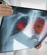
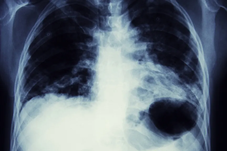
Typically a person presents with a past history or symptoms that warrant investigation with a chest x-ray. The chest x-ray is read as “abnormal” by the radiologist, who then usually states that a mass cannot be ruled out.
第一步是计算机断层扫描(CT)can of the chest. The purpose of the CT scan is to:
确认这确实这是一个坚实的质量,而不是气道或肺炎的融合。
衡量质量是多大的,识别其特征:平滑边界,不规则,推测等。其中一些特征将增加这是恶性肿块的可能性。
评估是否存在这种质量的延伸,仅仅是肺部的一个叶片,或者如果肺部外部延伸。
评估是否存在淋巴结显示出来。这将对癌症的扩散或转移产生影响。
所有实用目的肺癌are divided into two groups. One group is小细胞癌。所有其他肺癌类型都被共同称为非小细胞肺癌(NSCLC)。
如果它仅在一个肺部和“延长”之外,小细胞癌被归类为“有限”,如果它在相关的肺部外面蔓延。非小细胞癌通过更复杂的系统“阶段”。
通过国际肺癌研究协会建立了TNM(肿瘤,节点,转移)分期系统。从那时起,它已经修改并更新了八次,最新版本将于2018年1月实施。
分类系统考虑到:
这弥撒的大小(t),T1小于3厘米,T2大于3厘米。
淋巴结参与(N). Lymph nodes are connected to lymphatic channels and cancer involvement, when seen in the lymph nodes, implies that cancer cells have the leewayto spread to distant sites in the body (metastasis). This part of the classification system takes into account if the lymph nodes involved are within the lungs (hilar), or located in the chest wall between the lungs (mediastinal), or if the involved lymph nodes are outside the chest in areas like the neck or axillae (armpit).
转移(M) which identifies whether the cancer has been contained within the same lung (M0) or spread to areas outside the lungs (M1)
所有这些数据都是协调的,并导致I,II,III,IV的复杂分期系统,然后进一步细分一些阶段进入A,B,C。分类系统的原因是帮助临床医生确定每个患者肺癌的预后和治疗方案。通常,暂存至IIIa分类被认为是“通过手术切除的敏捷”。
这next step is Positron Emission Tomography (PET scan). A radioactive glucose “tracer” that allows visualization on scan is injected and then a special scan is done to determine the sites of the body that use the glucose in excess of neighboring areas. This excess metabolic activity (measured in SUV units) implies cancer activity and helps to identify if there is spread of the cancer cells.
接下来,患者将经历肺功能测试(PFT)。该测试评估患者的呼吸状态,以确定他是否能够承受肿瘤切除。同样考虑的是,一些呼吸功能差的一些患者可能无法耐受放射治疗,因为这种治疗可能导致肺部的瘢痕(这将在已经受损的患者中进一步降低呼吸状态)。在最后一刻,外科医生也必须具有能力,如有必要,请评估呼吸状况至关重要。该测试还将有助于预测手术后的呼吸状态(如果执行)。
考虑这些步骤初步测试。为了使肺癌正式诊断,直接从肿块中接受组织的活组织检查和对鉴定的细胞类型的病理报告确定是否存在有非典型的细胞表明癌症。
这活检通过几种方法中的一种完成:
一种支气管镜(fiber-optic tube) is inserted through the nose and through the airways of the lungs. When the location of the tumor is reached (determined by a real time x-ray called fluoroscopy), a forceps wire is inserted through the bronchoscope and a piece of tissue (biopsy) is obtained.
胚胎超声(ebus)引导活组织检查:如果肿瘤非常小或支气管内腔,则实时超声将有助于引导钳子的活组织检查捕获。
When the lesion is at the edge of the lung (where the bronchoscope will not reach), a needle is passed through the outer chest wall to obtain the biopsy, with the help of a CT scan. This is called a Percutaneous Needle Biopsy (PNB).
Video-assisted thorascopic surgery (VATS): In difficult cases, a last resort effort by a thoracic surgeon (requires general anesthesia) introduces a small video camera into the patient’s chest via a scope.
Next:选择最好的治疗