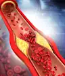
What Does Xanthelasma Have to Do With Cholesterol?
"Xanthos" means "yellow"in Greek. Xenthelasma are indeed skin deposits of yellow-colored fatty material and debris, the remnants of inflammatory cells (macrophages) bloated with lipid material that confers the yellow color. LDL particles are trapped in tissues, in this case in the connective tissue of the eyelids. Interestingly, the LDL particles trapped within the xanthelasma are oxidized, the form that also causes atherosclerotic plaque. That's the rub.
所以黄斑瘤不仅仅是curiosities or blemishes. Like many phenomena on skin, xanthelasma are the surface expression of an internal process.
But before they have a chance to form atherosclerotic plaque in coronary, carotid, and other arteries, the lipoproteins that eventually lead to xanthelasma can be identified as abnormalities in the blood. It can show up as very high LDL cholesterol values, often in the 250 mg/dl or more range, called "familial heterozygous hypercholesterolemia," an inherited disorder in which an LDL receptor is lacking or dysfunctional and LDL particles accumulate. Xanthelasma have also been associated with high levels of triglyceride-rich VLDL (very low-density lipoproteins), low HDL, and sometimes with no disorder of lipoproteins (at least not one that fits neatly into the usual categories).
Having xanthelasma should therefore prompt your doctor to ask whether there is a disorder of lipoproteins present that allows such a surface "red flag" to develop.
Having seen many people with such surface markers of lipoprotein disorders, I also have a hunch, one not yet supported by formal study, but one that fits nicely into the range of lipoprotein disorders commonly associated with xanthelasma. I suspect that xanthelasma are really the accumulation of小LDL particles, the abnormality that has leapt to first place as most common cause for heart disease in the U.S., the sort triggered by overconsumption of carbohydrates. Small LDL particles are the most adherent to structural tissues, such as those in vessels and skin, and the form most oxidation-prone. Small LDL is much more likely to be present when VLDL levels are high and when HDL levels are low. High (total) LDL levels are typically accompanied by some proportion of small LDL particles, the kind triggered by an apparently healthy diet filled, for example, with "healthy whole grains." (Grains are flagrant triggers of small LDL particles, contrary to conventional advice to eat more.)
The bottom line: If you or someone you know has little visible yellow plaques on their eyelids, an investigation should be made for a disorder of lipids or lipoproteins. In particular, small LDL particles should be measured along with total LDL, triglycerides, and HDL.
Even better, a direct measure of coronary risk should be considered, e.g., a CT heart scan, the low-radiation "mammogram"-equivalent to uncover coronary atherosclerotic plaque. Having either a significant lipoprotein abnormality and/or coronary plaque identified by a heart scan should then prompt an intensified effort to prevent the heart attack that is likely in future without proper preventive action.